PREIMPLANTATION GENETIC DIAGNOSIS FOR BRCA 1/2 CARRIERS
A
Assisted Embryo Hatching
a procedure in which a hole is made in the zona pellucida of the early embryo. The embryo may more easily “hatch” out of the zona through the hole prior to implantation.
Assisted Reproductive Technologies (ART)
a collective term which refers to a variety of medical procedures used to achieve pregnancy.
B
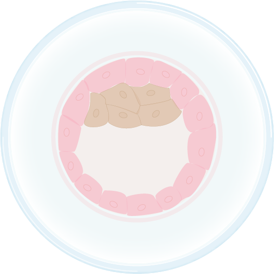
Blastocyst
the stage of development that the embryo is in when it enters the uterine cavity for implantation (typically 5-6 days after fertilization). It is comprised of two types of cells; an outer layer of cells called the trophoblast that will become the supporting tissue of the embryo such as the placenta and an inner layer referred to as the Inner Cell Mass, which will be multiply and contribute to the cells of the embryo.
Blastomere
a single cell from the developing 3-day embryo.
C
Cryopreservation
halting the embryo development before implantation through a freezing procedure.
Controlled Ovarian Hyperstimulation (COH)
the use of medications to stimulate growth and development of multiple ovarian follicles.
F
Follicle
a structure in the ovary that has nurtured the ripening egg and from which the egg is released or retrieved.
Follicle Stimulating Hormone (FSH)
a hormone produced by the pituitary gland that stimulates the ovary to ripen a follicle for ovulation.
Fluorescent in-situ hybridization (FISH)
a type of visualization used to determine the chromosome number and chromosomal mutation status of the embryo via PGD or PND.
G
Genetic counselor
a medical professional with specialized training in clinical genetics.
I
Intracytoplasmic Sperm Injection (ICSI)
an assisted fertilization technique in which a sperm is micro injected directly into the egg cytoplasm.
In Vitro Fertilization (IVF)
a procedure during which an egg is removed from a ripe follicle and fertilized by a sperm outside the human body.
L
Linkage Analysis
involves the study of BRCA1/2 inheritance patterns within a family and the identification of genetic markers that map near the gene. These genetic markers are physically linked to the gene.
M
Meiosis
this term describes cell division that occurs during the formation of the mature egg and sperm. During meiosis, the number of chromosomes (genes) is reduced so that the egg and sperm contribute only half of the parent’s genes to the embryo.
O
Ovarian Hyperstimulation Stimulation Syndrome (OHSS)
a medical complication of the stimulation procedures and medications in which stimulated follicles produce excess hormones and other factors which may lead to serious complications and may cause cancellation of a cycle and for a small number of women may result in death.
P
Polar body
a polar body is a small cell that is naturally released by the egg or oocyte during the process of meiosis. The first polar body is released by the oocyte near the time of ovulation. The second polar body is released by the oocyte, at the time of fertilization. The polar bodies do not contribute to the developing embryo, but are naturally discarded by the oocyte during the process of meiosis. Because the polar body contains one half of the possible genetic information of the mother, the genetic information of the oocyte can be inferred by the genetic testing of the polar body.
Polymerase chain reaction (PCR)
one of the most widely used laboratory techniques for visualizing genetic information and can be used for PGD or PND.
Preimplantation Genetic Diagnosis (PGD)
refers to procedures that are performed on embryos prior to implantation, sometimes even on oocytes prior to fertilization to determine gene status.
Prenatal Diagnosis (PND)
testing for genetic mutations in a fetus during the first or second trimester of pregnancy.
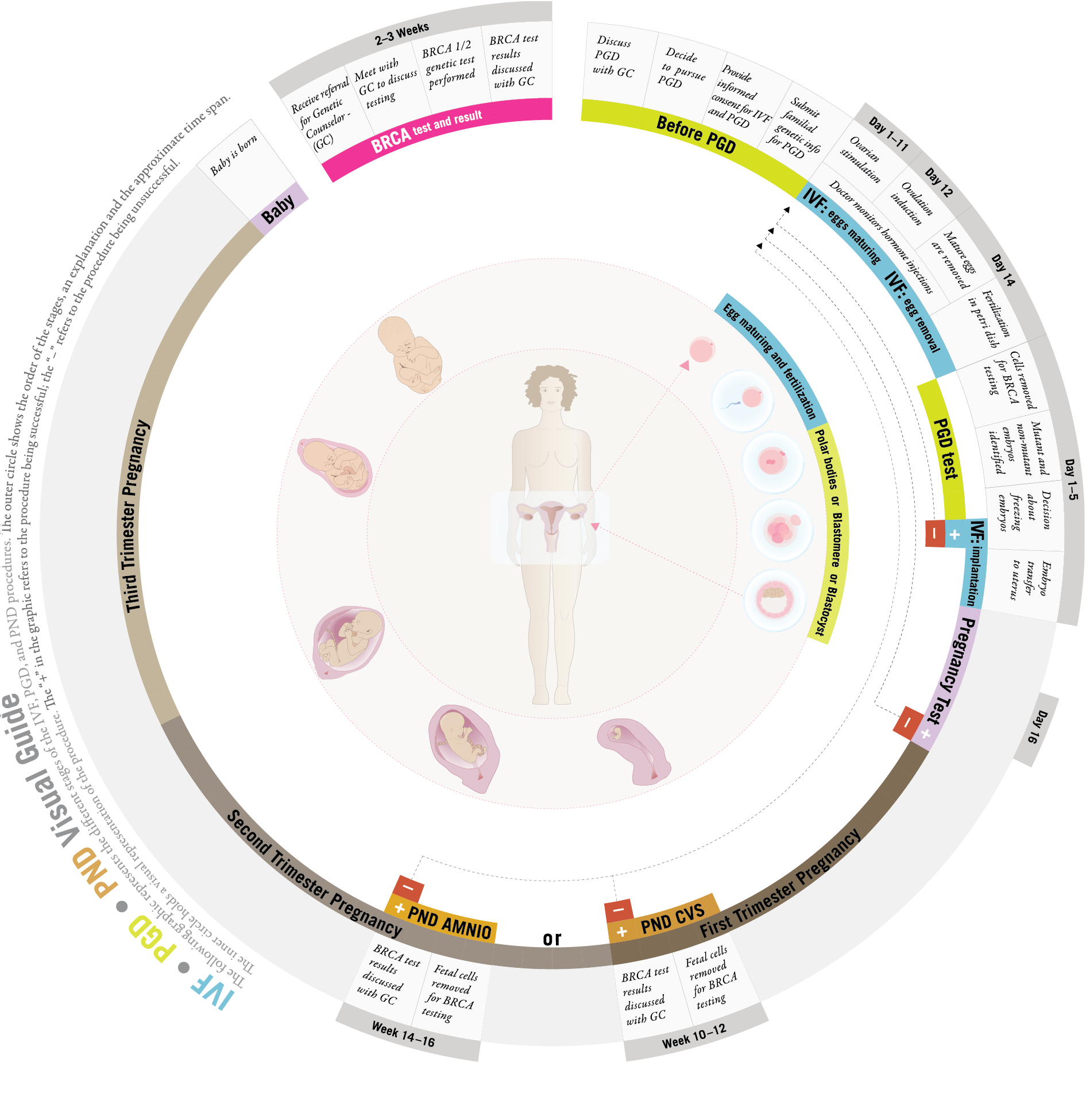
A Visual Guide to BRCA and PGD:
The following graphic represents the different stages of the IVF, PGD, and PND procedures. The outer circle shows the order of the stages, an explanation and the approximate time span. The inner circle holds a visual representation of the procedure.
The "+" in the graphic refers to the procedure being successful; the "–" refers to the procedure being unsuccessful.
Genetic Testing Methods:
Testing Options
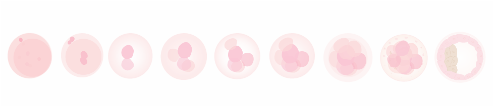
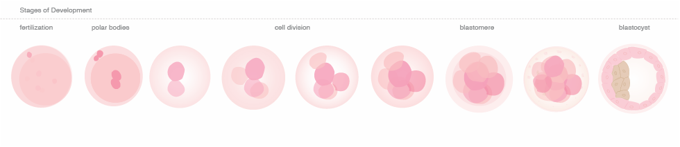
After the egg has been fertilized, PGD can be performed. PGD involves the removal of cellular material and the subsequent testing of the DNA in that material. As PGD techniques have advanced, clinicians can conduct the genetic testing at various time points post-fertilization. Depending on when genetic analysis is conducted, the number of cells removed may change. Today PGD can be conducted at one of three stages of embryonic development and clinics vary with regard to the timing and type of genetic testing.
PGD can be performed at:
Day 1 Polar Bodies – by the removal of a polar body, which is a byproduct of the first cell division post fertilization.

Day 3 Blastomere (8 cells) – by the removal of 1-2 cells of the early embryo

Day 5 Blastocyst (150 cells) – by the removal of multiple cells from the the outer layer of the embryo called the trophoblast, which will go on to become the cells of the placenta, while the inner cells go on to become the embryo.
Types of genetic visualization for PGD
Since the amount of genetic material that is taken from the embryo is a very small quantity, visualization techniques have been developed that allow detailed genetic analysis. Polymerase Chain Reaction (PCR) is a laboratory technique that amplifies a small amount of DNA from a single cell or small number of cells and allows for genetic testing outside of the cells. For dominant mutations like BRCA1/2, it is important to test two cells from the embryo using PCR. Another type of genetic visualization is Fluorescent in-situ hybridization (FISH), a technique commonly used to detect chromosomal abnormalities (eg. Trisomy 21). FISH involves the binding of specific DNA probes to regions of chromosomes in the cells.